Home
/ Neck And Shoulder Anatomy Diagram - Shoulder Head And Neck Anatomy Human Anatomy Png 600x600px Watercolor Cartoon Flower Frame Heart Download Free : Browse 3,107 anatomy of neck and shoulder stock photos and images available, or start a new search to explore more stock photos and images.
Neck And Shoulder Anatomy Diagram - Shoulder Head And Neck Anatomy Human Anatomy Png 600x600px Watercolor Cartoon Flower Frame Heart Download Free : Browse 3,107 anatomy of neck and shoulder stock photos and images available, or start a new search to explore more stock photos and images.
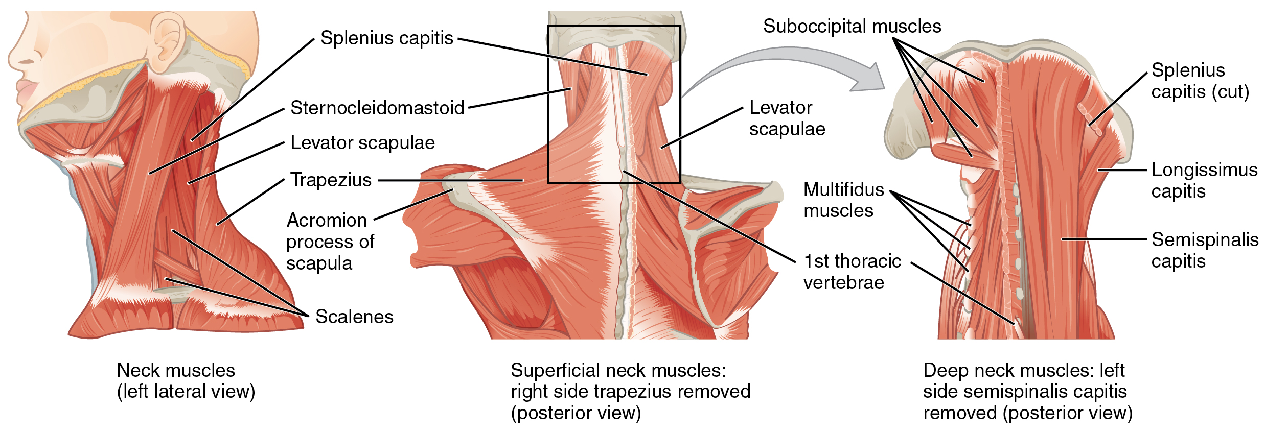
Neck And Shoulder Anatomy Diagram - Shoulder Head And Neck Anatomy Human Anatomy Png 600x600px Watercolor Cartoon Flower Frame Heart Download Free : Browse 3,107 anatomy of neck and shoulder stock photos and images available, or start a new search to explore more stock photos and images.. The neck muscles, including the sternocleidomastoid and the trapezius, are responsible for the gross motor movement in the muscular system of the head and neck. The anatomy of the neck and shoulders is very interesting. Contain the common carotid artery, internal. Although anchored in the neck, their primary functions are to move the shoulder blades and support the arms. Thank you for your support.
Pain in a man's body pain in a man's body on a gray background. Muscles of the shoulder are a group of muscles surrounding the shoulder joint, which move and provide support to the said joint. Two joints are at the shoulder. The shoulder joint is the junction between the chest and the upper extremity. Anterior, lateral and posterior groups, based on their position in the neck.the musculature of the neck is further divided into more specific groups based.
The Ventral Neck Muscles Lecturio Online Medical Library from d3uigcfkiiww0g.cloudfront.net The content of the neck is grouped into 4 neck spaces, called the compartments. 🤔 the acetabulofemoral joint , commonly called the hip joint , scientifically termed is located in between the pelvis and the femur of the legs. The neck muscles, including the sternocleidomastoid and the trapezius, are responsible for the gross motor movement in the muscular system of the head and neck. There are eight pairs of cervical nerves, denoted c1 to c8. The humerus or one of the other bones in the shoulder slips out of position. Although anchored in the neck, their primary functions are to move the shoulder blades and support the arms. Anterior, lateral and posterior groups, based on their position in the neck.the musculature of the neck is further divided into more specific groups based. They move the head in every direction, pulling the skull and jaw towards the shoulders, spine, and scapula.
The cervical spine is responsible for several crucial roles, including.
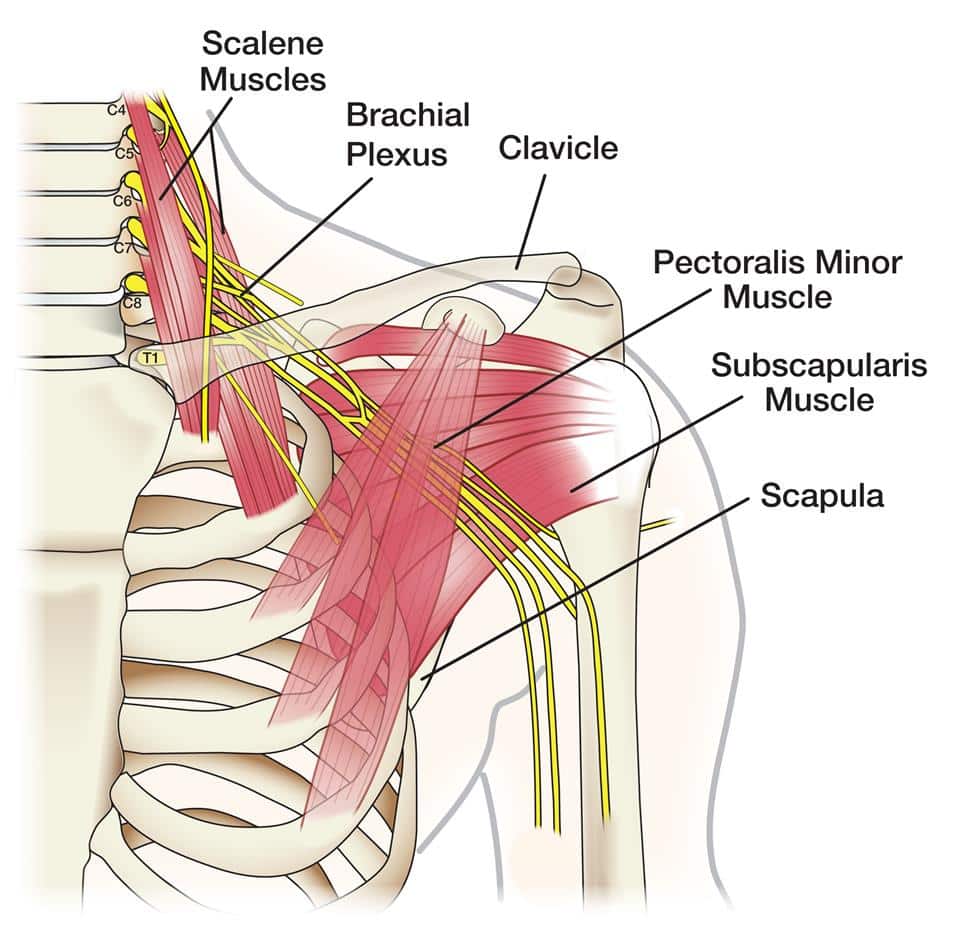
While classified as peripheral nerves, the motor cell body resides in the anterior horn of the spinal cord. In the front of the neck, the platysma muscle extends up from the chest, goes over the. Bones have many shapes and sizes and are important to add structure to the body and protection to the the shoulder girdle combines to give you shoulder motion. The muscles of the shoulder are associated with movements at the shoulder joint. These nerves conduct motor and sensory information via efferent and afferent fibers, respectively, to and from the central nervous system. The humerus or one of the other bones in the shoulder slips out of position. The cervical spine is responsible for several crucial roles, including. The majority of these nerves control the functions of the upper extremities and allow you to feel your arms, shoulder, and back of your head. Interactive anatomical atlas of the head, brain, and neck based on anatomical diagrams and ct and mri medical imaging exams. Anatomy if neck and back diagram. Rectus capitis posterior major, which arises from the spinous process of the axis (c2). Neck and shoulder pain anatomy. These are the supraspinatus, infraspinatus, teres.
Interactive anatomical atlas of the head, brain, and neck based on anatomical diagrams and ct and mri medical imaging exams. 🤔 the acetabulofemoral joint , commonly called the hip joint , scientifically termed is located in between the pelvis and the femur of the legs. Although anchored in the neck, their primary functions are to move the shoulder blades and support the arms. In human anatomy the shoulder joint comprises the part of the body where the humerus attaches to the scapula the head sitting in the glenoid cavity. Anatomy if neck and back diagram.
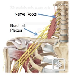
Neck Pain Referred To The Shoulder Shoulderdoc from www.shoulderdoc.co.uk These are the supraspinatus, infraspinatus, teres. Each nerve provides sensation to a specific area of the body called a dermatome. Human shoulder anatomy posterior view of the shoulder anatomy pinterest shoulder. The clavicle is the only bony attachment between the trunk and the upper limb. Rectus capitis posterior major, which arises from the spinous process of the axis (c2). In human anatomy the shoulder joint comprises the part of the body where the humerus attaches to the scapula the head sitting in the glenoid cavity. The shoulder is one of the largest and most complex joints in the body. Browse 3,107 anatomy of neck and shoulder stock photos and images available, or start a new search to explore more stock photos and images.
The shoulder joint is the junction between the chest and the upper extremity.
They move the head in every direction, pulling the skull and jaw towards the shoulders, spine, and scapula. The shoulder joint is the junction between the chest and the upper extremity. Arm and shoulder bones the upper arm bone called the humerus is connected to the body via the shoulder blade which possesses the latin name scapula. The muscles of the shoulder are associated with movements at the shoulder joint. Contains cervical vertebrae and postural muscles. Each nerve provides sensation to a specific area of the body called a dermatome. The neck and shoulders are complex and interconnected areas, and medical problems that affect one often affect the other, as well. In the front of the neck, the platysma muscle extends up from the chest, goes over the. Neck neck muscle anatomy muscle diagram inspirational medical. These critical parts of the upper body are very prone to developing pain because the position of all the bones in the neck and shoulders are completely dependent on the balance and alignment of the muscles and fascia that lash them together and allow for movement between them. Rectus capitis posterior major, which arises from the spinous process of the axis (c2). The anatomy of the neck and shoulders is very interesting. The muscles of the neck run from the base of the skull to the upper back and work together to bend the head and.
Human shoulder anatomy posterior view of the shoulder anatomy pinterest shoulder. Veins and arteries of the neck 9 photos of the veins and arteries of the neck activate javascript arteries in the neck diagram, common carotid artery branches, external carotid artery function, how many carotid arteries, left common carotid artery function, the left common carotid artery supplies blood to the. Cat anatomy dissection guide superficial muscles ventral view pectoantebrachialis dorsal view clavotrapezius. In human anatomy the shoulder joint comprises the part of the body where the humerus attaches to the scapula the head sitting in the glenoid cavity. The back's muscles start at the top of the back (named the cervical vertebrae) and go to the tailbone (also named the coccyx).
Shoulder Anatomy Elliot S Site from elliottelford.com The majority of these nerves control the functions of the upper extremities and allow you to feel your arms, shoulder, and back of your head. These consist of the arm, located between the shoulder and elbow joints; 🤔 the acetabulofemoral joint , commonly called the hip joint , scientifically termed is located in between the pelvis and the femur of the legs. These critical parts of the upper body are very prone to developing pain because the position of all the bones in the neck and shoulders are completely dependent on the balance and alignment of the muscles and fascia that lash them together and allow for movement between them. These are the supraspinatus, infraspinatus, teres. Begin at the ear and work down to where the neck meets the shoulder. Numerous muscles help stabilize the three joints of. Contains cervical vertebrae and postural muscles.
These are the supraspinatus, infraspinatus, teres.
Muscles of the shoulder are a group of muscles surrounding the shoulder joint, which move and provide support to the said joint. Contains glands ( thyroid, parathyroid, and thymus ), the larynx, pharynx and trachea. Pain and dysfunction from injuries or conditions that impact the joints, muscles, and other structures can easily spread from the neck to the shoulder(s) and from the shoulder(s) to the neck. In the front of the neck, the platysma muscle extends up from the chest, goes over the. The upper arm bone, called the humerus, is connected to the body via the shoulder blade, which possesses the latin name scapula. There are eight pairs of cervical nerves, denoted c1 to c8. The shoulder joint also known as the glenohumeral joint is the main joint of the shoulder. The neck muscles, including the sternocleidomastoid and the trapezius, are responsible for the gross motor movement in the muscular system of the head and neck. A second joint in the shoulder is the junction of the collar bone with the shoulder blade, called the. Muscles of the neck (musculi cervicales) the muscles of the neck are muscles that cover the area of the neck hese muscles are mainly responsible for the movement of the head in all directions they consist of 3 main groups of muscles: Begin at the ear and work down to where the neck meets the shoulder. Contains cervical vertebrae and postural muscles. These critical parts of the upper body are very prone to developing pain because the position of all the bones in the neck and shoulders are completely dependent on the balance and alignment of the muscles and fascia that lash them together and allow for movement between them.
🤔 the acetabulofemoral joint , commonly called the hip joint , scientifically termed is located in between the pelvis and the femur of the legs shoulder anatomy diagram. There are 8 spinal nerves that originate from the cervical spine.